Muscles Labeled Front And Back / Muscle Labeling - Anatomy with E at West Springfield High School - StudyBlue. Superficial muscles are the muscles closest to the skin surface and can usually be seen while a body is performing actions. Gluteus maximus, semitendinosus and biceps. Intermediate back muscles and c. Intermediate back muscles and c. Within this group of back muscles you will find the latissimus dorsi, the trapezius, levator scapulae and the rhomboids.
More specifically, this beautifully illustrated anatomy chart includes neck and shoulders, multiple views of the back and spine, and frontal views of each muscular extremity of the human body. Labeled viral infection explanation scheme. Click on the labels below to find out more about your muscles. Human muscle system, the muscles of the human body that work the skeletal system, that are under voluntary control, and that are concerned with movement, posture, and balance. Label the following anatomicalsites in the diagram:
Muscular System Gifts on Zazzle from rlv.zcache.com Back » skeleton labeled front and back and muscles naming skeletal muscles anatomy and physiology categories: It is responsible for extension,adduction, and (medial) internal rotation of the shoulder joint. Labeled medical scheme with humerus, muscle, radius and ulna isolated closeup. Muscles during both the front and the back squat. But statistically significant differences were seen for. The anterior muscles of the torso (trunk) are those on the front of the body, including the muscles of the chest, abdomen, and pelvis. Vector illustration informative medical scheme. Back of the head muscle structure and nerve system diagram.
It attaches to the clavicle and scapula.
This labeled human muscular system chart illustrates the major muscle groups in the back (posterior) view and the front (anterior) view. Intermediate back muscles and c. There are two parallel muscles. The muscles of the back that work together to support the spine, help keep the body upright and allow twist and bend in many directions. Liver inflammation with scar tissues and cirrhosis. Labeled educational inner organ structure. Related searches for muscle diagram labeled front muscle diagram front and backfront and back body diagramanterior muscle diagramlabel the back musclesbody diagram muscles labelmuscular system anterior diagramfront body anatomyanterior muscles labeled. Within this group of back muscles you will find the latissimus dorsi, the trapezius, levator scapulae and the rhomboids. Click on the labels below to find out more about your muscles. Triceps, biceps, pectoralis major, quadriceps , hamstrings, gluteus maximus , abdominals, deltoid, latissimus dorsi, external obliques, gastrocnemius , tibialis anterior. Back » skeleton labeled front and back and muscles naming skeletal muscles anatomy and physiology categories: This muscular system chart shows in detail the deep layers of muscle on the back side of your body. The biggest muscle is lats muscle, then there are traps muscle.
The muscles of the anterior of the forearm are generally divided into two groups: These muscles are able to move the upper limb as they originate at the vertebral column and insert onto. Intermediate back muscles and c. Labeled tick bite infection symptoms scheme. Back view of muscles, skeleton, organs, nervous system.
Muscles of the Pectoral Girdle and Upper Limbs | Anatomy and Physiology I from s3-us-west-2.amazonaws.com Triceps, biceps, pectoralis major, quadriceps , hamstrings, gluteus maximus , abdominals, deltoid, latissimus dorsi, external obliques, gastrocnemius , tibialis anterior. Labeled viral infection explanation scheme. The superficial back muscles are the muscles found just under the skin. Human muscle system, the muscles of the human body that work the skeletal system, that are under voluntary control, and that are concerned with movement, posture, and balance. These muscles are able to move the upper limb as they originate at the vertebral column and insert onto. Liver inflammation with scar tissues and cirrhosis. Labeled medical scheme with humerus, muscle, radius and ulna isolated closeup. This muscular system chart shows in detail the deep layers of muscle on the back side of your body.
More specifically, this beautifully illustrated anatomy chart includes neck and shoulders, multiple views of the back and spine, and frontal views of each muscular extremity of the human body.
Male muscular system, full anatomical body diagram with muscle scheme, vector illustration educational poster. Labeled viral infection explanation scheme. Virus disease symptoms and spreads infographic. Back » skeleton labeled front and back and muscles naming skeletal muscles anatomy and physiology categories: Muscle label thoracic region (back + front). More specifically, this beautifully illustrated anatomy chart includes neck and shoulders, multiple views of the back and spine, and frontal views of each muscular extremity of the human body. But statistically significant differences were seen for. The spinal cord, which controls over 10 billion nerve cells, is less than two feet in length and its diameter is same as that of the index finger. The muscles of the back that work together to support the spine, help keep the body upright and allow twist and bend in many directions. Leg muscle anatomical structure, labeled front, side and back view diagrams. Many in the neck help to stabilize or move the head. There are two parallel muscles. Back view of muscles, skeleton, organs, nervous system.
Liver inflammation with scar tissues and cirrhosis. Virus disease symptoms and spreads infographic. It is responsible for extension,adduction, and (medial) internal rotation of the shoulder joint. Back of the head muscle structure and nerve system diagram. The anterior muscles of the torso (trunk) are those on the front of the body, including the muscles of the chest, abdomen, and pelvis.
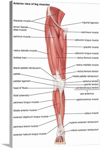
Muscles of the leg - anterior view Wall Art, Canvas Prints, Framed Prints, Wall Peels | Great ... from static.greatbigcanvas.com Virus disease symptoms and spreads infographic. Label muscles front and back view. These muscles are able to move the upper limb as they originate at the vertebral column and insert onto. Back view of muscles, skeleton, organs, nervous system. Many in the neck help to stabilize or move the head. It attaches to the clavicle and scapula. But statistically significant differences were seen for. Muscles during both the front and the back squat.
The spinal cord, which controls over 10 billion nerve cells, is less than two feet in length and its diameter is same as that of the index finger.
Virus disease symptoms and spreads infographic. The anterior muscles of the torso (trunk) are those on the front of the body, including the muscles of the chest, abdomen, and pelvis. Intermediate back muscles and c. Labeled medical scheme with humerus, muscle, radius and ulna isolated closeup. Aalso known as the six pack, is a paired muscle running vertically on each side of the front wall of the abdomen. Back view of muscles, skeleton, organs, nervous system. A back muscle that pulls the arm down and back. Labeled viral infection explanation scheme. Superficial muscles are the muscles closest to the skin surface and can usually be seen while a body is performing actions. But statistically significant differences were seen for. Labeled medical scheme with humerus, muscle, radius and ulna isolated closeup. Label muscles front and back view. Label muscles front and back view.
Share :
Post a Comment
for "Muscles Labeled Front And Back / Muscle Labeling - Anatomy with E at West Springfield High School - StudyBlue"


Post a Comment for "Muscles Labeled Front And Back / Muscle Labeling - Anatomy with E at West Springfield High School - StudyBlue"